
Exercise 17 brain and cranial nerves – Exercise 17: Brain and Cranial Nerves embarks on a comprehensive exploration of the intricate anatomy and functions of the brain and cranial nerves. This detailed guide provides a systematic approach to performing Exercise 17, equipping readers with a thorough understanding of the neurological examination and its significance in diagnosing neurological disorders.
Through an engaging narrative, this guide unravels the complex relationship between the brain and cranial nerves, highlighting their vital roles in sensory and motor functions. By delving into the clinical applications of Exercise 17, readers gain valuable insights into the identification and localization of neurological deficits, empowering them to make informed clinical decisions.
Exercise 17: Brain and Cranial Nerves: Exercise 17 Brain And Cranial Nerves
Exercise 17 is a comprehensive neurological examination that assesses the function of the cranial nerves and the brainstem. It is an essential component of a thorough neurological examination and provides valuable information about the integrity of the nervous system.
The anatomical structures involved in Exercise 17 include the cranial nerves, brainstem, and associated structures such as the eyes, ears, nose, and mouth. The cranial nerves are responsible for transmitting sensory and motor information between the brain and various parts of the head and neck.
Step-by-Step Guide to Performing Exercise 17
- Inspect the patient’s face for symmetry and any signs of drooping or weakness.
- Test the sense of smell by asking the patient to identify different scents.
- Check the visual fields by asking the patient to follow a moving object with their eyes.
- Assess pupillary reflexes by shining a light in the patient’s eyes.
- Test extraocular movements by asking the patient to look in different directions.
- Evaluate the corneal reflex by touching the patient’s cornea with a wisp of cotton.
- Test the gag reflex by stimulating the back of the patient’s throat.
- Check the palate for symmetry and movement.
- Assess the tongue for movement and strength.
- Test the sternocleidomastoid and trapezius muscles for strength.
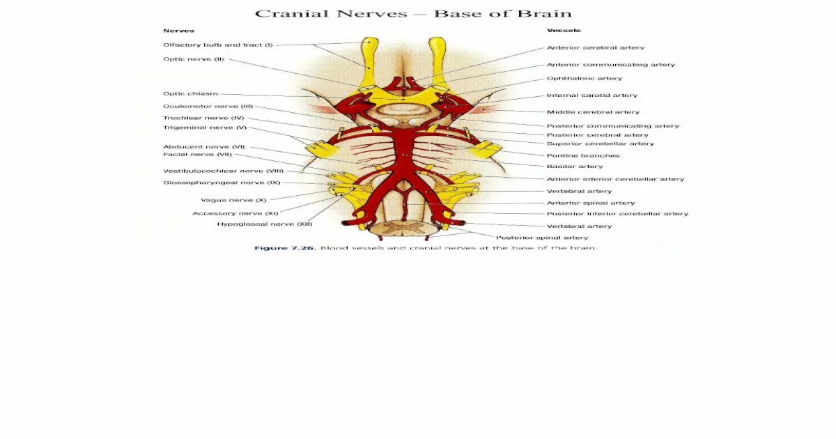
Cranial Nerves
The 12 cranial nerves are:
- Olfactory nerve (CN I): Smell
- Optic nerve (CN II): Vision
- Oculomotor nerve (CN III): Eye movement
- Trochlear nerve (CN IV): Eye movement
- Trigeminal nerve (CN V): Sensation and motor function of the face
- Abducens nerve (CN VI): Eye movement
- Facial nerve (CN VII): Facial expression
- Vestibulocochlear nerve (CN VIII): Hearing and balance
- Glossopharyngeal nerve (CN IX): Taste and swallowing
- Vagus nerve (CN X): Swallowing, speech, and heart rate
- Accessory nerve (CN XI): Neck and shoulder movement
- Hypoglossal nerve (CN XII): Tongue movement
Clinical Significance of Testing Cranial Nerves
Testing cranial nerves is essential for detecting neurological deficits and localizing lesions within the brainstem and surrounding structures. Abnormalities in cranial nerve function can indicate damage to the nerve itself, its nucleus, or the pathways connecting them.
| Cranial Nerve | Function | Associated Tests |
|---|---|---|
| Olfactory (CN I) | Smell | Identify different scents |
| Optic (CN II) | Vision | Visual field testing, pupillary reflexes |
| Oculomotor (CN III) | Eye movement | Extraocular movements, pupillary reflexes |
| Trochlear (CN IV) | Eye movement | Extraocular movements |
| Trigeminal (CN V) | Sensation and motor function of the face | Sensory testing, motor function testing |
| Abducens (CN VI) | Eye movement | Extraocular movements |
| Facial (CN VII) | Facial expression | Facial symmetry, movement testing |
| Vestibulocochlear (CN VIII) | Hearing and balance | Hearing tests, balance testing |
| Glossopharyngeal (CN IX) | Taste and swallowing | Taste testing, gag reflex |
| Vagus (CN X) | Swallowing, speech, and heart rate | Gag reflex, palatal movement, speech testing |
| Accessory (CN XI) | Neck and shoulder movement | Sternocleidomastoid and trapezius muscle testing |
| Hypoglossal (CN XII) | Tongue movement | Tongue movement testing |
Neurological Examination
Exercise 17 is a key component of a comprehensive neurological examination. It provides information about the function of the cranial nerves and brainstem, which is essential for diagnosing a wide range of neurological disorders.
Components of a Neurological Examination, Exercise 17 brain and cranial nerves
- Mental status examination
- Cranial nerve examination (Exercise 17)
- Motor examination
- Sensory examination
- Reflex examination
- Gait examination
Importance of a Thorough Neurological Examination
A thorough neurological examination is essential for diagnosing neurological disorders because it allows the examiner to:
- Detect neurological deficits
- Localize lesions within the nervous system
- Monitor disease progression
- Evaluate treatment response
Case Studies
Case Study 1:A 65-year-old woman presents with sudden onset of right-sided facial droop and difficulty swallowing. Exercise 17 reveals weakness of the right facial muscles, impaired gag reflex, and deviation of the uvula to the left. These findings suggest a lesion involving the right facial nerve (CN VII) and the right vagus nerve (CN X).
Case Study 2:A 40-year-old man presents with progressive weakness of the left arm and leg. Exercise 17 reveals weakness of the left facial muscles, impaired hearing on the left side, and decreased sensation to touch on the left side of the body.
These findings suggest a lesion involving the left facial nerve (CN VII), the left vestibulocochlear nerve (CN VIII), and the left trigeminal nerve (CN V).
These case studies demonstrate how Exercise 17 findings can help identify and localize neurological deficits. By assessing the function of the cranial nerves, clinicians can gain valuable information about the location and extent of neurological damage.
Advanced Techniques

In addition to the basic techniques described in Exercise 17, there are a number of advanced techniques that can be used to assess cranial nerve function.
Electromyography and Nerve Conduction Studies
Electromyography (EMG) and nerve conduction studies (NCS) are electrodiagnostic tests that can be used to evaluate the function of individual nerves and muscles. EMG measures the electrical activity of muscles, while NCS measures the conduction of electrical impulses along nerves.
Imaging Studies
Imaging studies, such as magnetic resonance imaging (MRI) and computed tomography (CT), can be used to visualize the cranial nerves and surrounding structures. These studies can help identify structural abnormalities that may be causing cranial nerve dysfunction.
Questions and Answers
What is the purpose of Exercise 17?
Exercise 17 is a comprehensive neurological examination that evaluates the function of the brain and cranial nerves, providing insights into their anatomical and physiological integrity.
How many cranial nerves are there, and what are their functions?
There are 12 pairs of cranial nerves, each with specific sensory, motor, or autonomic functions. They play crucial roles in vision, hearing, smell, taste, facial movement, and other vital bodily processes.
What is the clinical significance of testing cranial nerves?
Testing cranial nerves during Exercise 17 helps identify deficits in sensory, motor, or autonomic function, which can indicate damage to specific cranial nerves or the structures they innervate. This information aids in the diagnosis and localization of neurological disorders.
